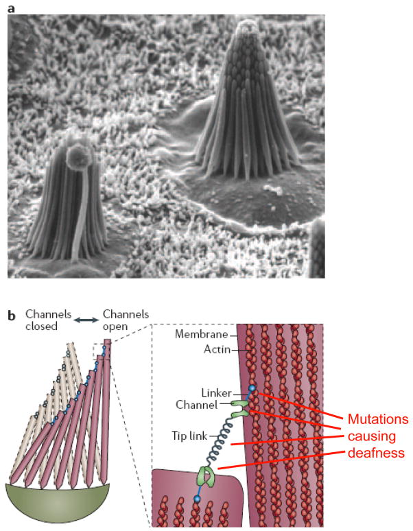
Figure 1. Mechanotransduction in hair cells.
(a). Scanning electron micrograph of two hair bundles in the sensory macula of the bull frog saccule, showing the arrangement of stereocilia with increasing heights. These bundles are ~8μm tall and contain 50–60 stereocilia. (b) Schematic drawing of a hair bundle in resting (gray) and deflected (color) configuration. Deflection, i.e., shearing of the stereocilia relative to each other, causes the ~150–200nm long tip links to pull directly on K+ channels in the stereocilia, causing the channels to open. Myosin motors that link the channels to the actin core of the sterocilia can adjust the position to restore resting tension in the tip link, allowing adaptation to persistent stimulation. Mutations in the K+ channel, the linker proteins, or the unconventional myosins (UCM), which keep the tip links under tension, can result in deafness. Figure is modified from Ref (82). Part a of the Figure is reproduced and part b of the Figure is modified with permission from Ref. (83)

