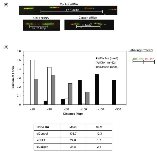
Figure 2. Chk1 and Claspin regulate replication origin density.
(A) Representative images of DNA fibers prepared from Chk1, Claspin or control siRNA-transfected cells pulsed with 25μM BrdU for 20 minutes followed by 250μM IdU for 20 minutes. Pattern of red-green staining shows the direction of synthesis in adjacent replication forks and allows determination of initiation sites such that inter-origin distance (I) can be measured. (B) Graph represents distribution of inter-origin distances in cells expressing control, Chk1 or Claspin siRNA. Table summarizes mean and standard error of the mean for distances measured from five independent experiments.

