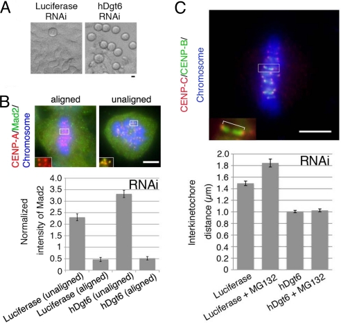
Fig. 4.
Reduced metaphase kinetochore tension in the absence of human augmin. (A) Mitotic cells (rounded cells in the phase-contrast microscopy image) were accumulated after hDgt6 RNAi. (B) The average intensity of the kinetochore-localized Mad2 signals normalized to that of CENP-A (n ≥ 41 kinetochores from ≥3 cells each). The Mad2 signals diminished on the aligned kinetochores in both the control and hDgt6-depleted cells. The brightest kinetochore Mad2 signal in each cell was also comparable between RNAi and control samples (Fig. S3C). (C) Reduced tension on sister kinetochores after hDgt6 RNAi. (Lower) The interkinetochore distance of the congressed chromosomes (blue) was quantified based on immunostaining of CENP-B (green) and CENP-C (red) (≥20 pairs of kinetochores from ≥6 cells each). (Upper) An enlarged image of a pair of sister kinetochores. Treatment with the proteasome inhibitor MG132 (10 μM, 90 min), which prolongs metaphase, did not alter the interkinetochore distance of hDgt6-depleted cells. (Scale bars, 5 μm.)

