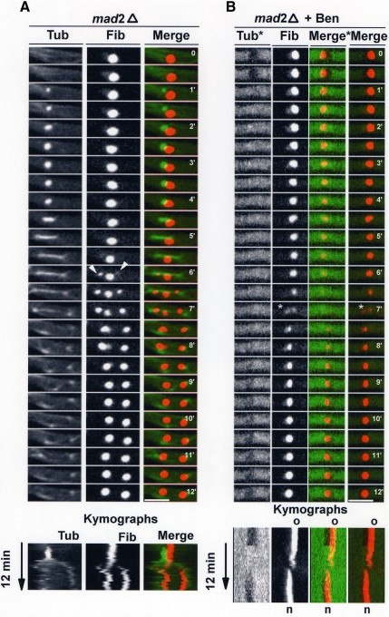
Figure 6.
Nucleolar segregation and the mitotic spindle. (A) Time-lapse images of mitosis in a mad2Δ strain containing GFP-tagged tubulin and An-Fib-chRFP. Arrowheads indicate the onset of daughter nucleoli formation, which occurs while the parental nucleolus is still present. (B) The same strain as in A undergoing a SIM in the presence of benomyl. In the Fib and Merge panels, the conditions and contrast are the same as in A. In the Tub* and Merge* panels, the contrast for tubulin has been adjusted to show the nuclear exclusion of depolymerized tubulin in interphase but not mitosis. A cycle of An-Fib disassembly from the parental nucleolus and reassembly at a distinct new nucleolus occurs (*) in the absence of a spindle. Kymographs are shown to highlight these changes. The period shown in the above montage is indicated on the kymographs. Bar, ∼5 μm.

