Figure 1.
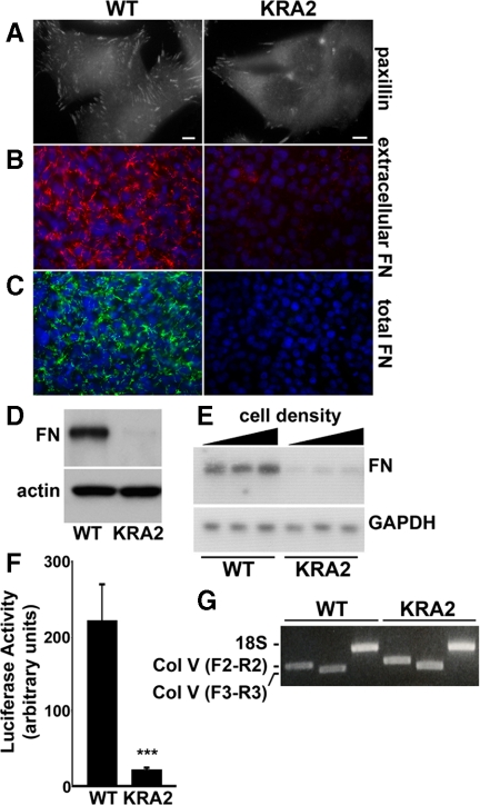
Fibronectin expression and transcription are decreased in fibroblasts expressing mislocalized NHE1-KRA2. (A) Paxillin immunolabeling reveals decreased abundance and size of FA complexes in KRA2 cells compared with WT cells. Bar, 5 μm. (B) Live-cell immunolabeling with FN antibodies reveals a marked decrease in secreted FN in KRA2 cells compared with WT cells (red, extracellular FN; blue, Hoechst staining for nuclei). (C) Total FN in permeabilized cells is decreased in KRA2 cells compared with WT cells (green, total FN; blue, Hoechst staining for nuclei). (D) Immunoblotting culture medium indicates secreted FN is attenuated in KRA2 cells compared with WT cells. Immunoblotting for β-actin in cell lysates was used to confirm equivalent cell numbers used to measure secreted FN. (E) Abundance of FN mRNA in KRA2 cells is attenuated compared with WT cells as determined by Northern blotting with GAPDH used as a loading control. FN transcription was not affected by cell density and was similar with 25, 50, and 95% confluency. Data are representative of blots from mRNA isolated from three separate cell preparations. (F) FN promoter activity, determined using a fragment of the human FN promoter between −105 and +14, is markedly decreased in the KRA2 cells compared with WT cells. Data represent means ± SEM of three separate cell preparations; *significant difference compared with WT; p < 0.001. (G) RT-PCR showing transcripts for hamster collagen α1 (V) chain in WT and KRA2 fibroblasts. Indicated are products using collagen primers F2 to R2 and F3 to R3, as described in Materials and Methods, and products of control primers for 18S RNA. Data are representative of two independent cell preparations.

