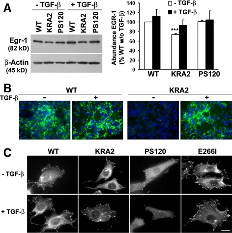
Figure 6.
TGF-β1 increases Egr-1 expression and restores a secreted and assembled FN matrix and the abundance of FA in KRA2 cells. (A) Representative immunoblot (left) and means ± SEM of data from immunoblots of three separate cell preparations (right) indicate that expression of Egr-1 is decreased in KRA2 cells compared with WT and PS120 cells. Incubation with (rh)TGF-β1 (5 ng/ml for 48 h) increases Egr-1 expression in all three cell types. Immunoblotting for β-actin was used as a loading control. *Significant difference compared with WT and PS120 cells; p < 0.001. (B) FN production and assembly, although nearly absent in KRA2 cells, are similar to WT cells after treating with 5 ng/ml TGF-β for 48 h, as determined by immunolabeling for extracellular FN. Data are representative of three separate cell preparations. (C) FA, as determined by paxillin immunolabeling in the indicated cell types. Treating cells with rhTGF-β1 increases the abundance and size of FA in KRA2 cells but not in WT, PS120, or E266I cells. Bar, 10 μM.

