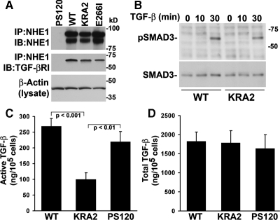
Figure 7.
Activation of TGF-β but not secretion of latent TGF-β or signaling by TGF-β-RI is inhibited in KRA2 cells. (A) TGF-β-RI coprecipitates with wild-type and mutant NHE1-HA. Immune complexes were immunoblotted with antibodies to NHE1, which recognize glycosylated (top band) and unglycosylated (bottom band) protein, and to TGF-β-RI. Aliquots of lysates were immunoblotted for β-actin to confirm equivalent amount of protein used for immunoprecipitation. Data are representative of two separate cell preparations. (B) Time-dependent increase in phosphorylated SMAD3 (pSMAD3) is similar in WT and KRA2 cells treated with 5 ng/ml rhTGF-β1. Immunoblotting for total SMAD3 indicates similar abundance in WT and KRA2 cells in the absence and presence of TGF-β. (C) Secreted active TGF-β determined by luciferase assays using MLEC cells expressing a truncated plasminogen activator inhibitor-1 (PAI-1) promoter and treated with conditioned medium from WT, KRA2, and PS120 cells. Luciferase units from MLEC cells treated with rhTGF-β1 were used to generate a standard curve and data, expressed as amount of TGF-β per 105 cells, represent means ± SEM of six cell preparations. (D) Secreted latent TGF-β determined as in C, but conditioned medium was incubated at 80°C for 10 min before adding to MLEC cells. Data are means ± SEM of four cell preparations.

