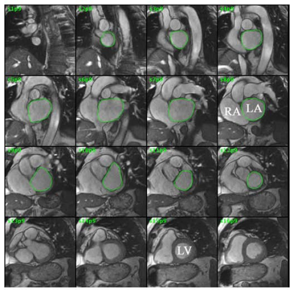
Figure 2.
MRI of left atrium. Steady state free precession cine gradient echo was performed on sequential slices of the left atrium. The left atrial border was traced at end-left ventricular systole. The summation of the areas multiplied by slice thickness is the left atrial volume. LA = left atrium, RA = right atrium, LV = left ventricle.

