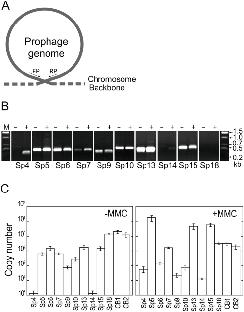
Figure 3. Excision, circularization, and replication of prophages.
(A) Schematic representation of the strategy to detect excised and circularized prophage genomes by PCR. (B) Detection of PCR products derived from excised and circularized prophage genomes by agarose gel electrophoresis. Total cellular DNA isolated from untreated (−) and MMC-treated cells (+) was analyzed. We examined all Sps, but only the data from the nine prophages that gave positive results are shown. The data of Sp18, a Mu-like phage whose DNA is not circularized, are shown as a negative control. (C) Quantification of circularized phage genomes in the total cellular DNA isolated from untreated (−MMC) and MMC-treated cells (+MMC) using quantitative PCR (qPCR). The data were obtained from three independent analyses, and average copy numbers for each prophage genome are shown. Bars indicate standard deviations. The Sp18 DNA and the chromosomal DNA from two chromosome regions (CB1 and CB2) were quantified as controls.

