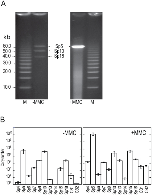
Figure 4. Phage DNA packaged into phage particles.
(A) FIGE analysis of packaged phage DNA in untreated (−MMC) and MMC-treated (+MMC) cultures. Phage particles were collected from culture supernatants by PEG/NaCl precipitation. DNA preparations applied to the FIGE gel were obtained from 50-ml (untreated) and 1-ml (MMC-treated) cultures. (B) Quantification of packaged phage DNA in the culture supernatants obtained from untreated (−MMC) and MMC-treated (+MMC) O157 Sakai cells. DNase-resistant phage DNA was quantified by qPCR using the primers that were used in Figure 3. DNA preparations equivalent to the 1-ml culture supernatant were used as template DNA. The data were obtained from three independent analyses, and the average copy numbers for each prophage genome are shown. Bars indicate standard deviations. The Sp18 DNA and the chromosomal DNA from two chromosome regions (CB1 and CB2) were monitored as controls.

