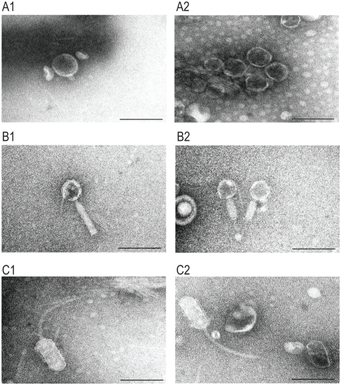
Figure 7. Electron micrographs of phage particles produced by O157 Sakai.
Electron micrographs of three types of phage particles detected in the culture supernatant of O157 Sakai are shown. (A) Phage particles with a short tail. (B) Phage particles with a contractile and non-flexible tail. (C) Phage particles with an elongated head and a long flexible tail. Phage particles shown in (A) are derived from Sp5 because K-12 strains lysogenized by CmR-marked Sp5 produced phage particles with the identical morphology. The similarity to phage Mu suggests that the phage particles shown in (B) are derived from Sp18. Electron micrographs were taken at magnifications of 60 K or 80 K using a transmission electron microscope and negatively contrasted with 2% uranyl acetate dihydrate. Bar, 100 nm.

