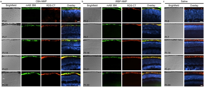
Figure 2. Transferred NMP co-localizes with endogenous RDS.
Frozen retinal sections from eyes collected at multiple ages (PI-2 to PI-30) were immunostained for NMP (mAB 3B6, green) and total RDS (RDS-CT, red) with a nuclear counterstain (DAPI, blue). Transferred RDS from eyes injected with CBA-NMP (A) and IRBP-NMP (B) nanoparticles is detected at PI-2. Expression remains strong through the latest time point analyzed (PI-30) and co-localizes with native RDS. Expression is limited to the OSs or nascent OSs and is not detected in any other retinal cell types, subcellular compartments or layers. (C) No NMP is detected in saline-injected control eyes, but native RDS is detected beginning at PI-2 (P7), consistent with normal ocular development. Scale bars, 20 µm. N = 3–5 mice per group. Abbreviations: RPE, retinal pigment epithelium; OS, outer segment layer; ONL, outer nuclear layer; INL, inner nuclear layer.

