FIG. 5.
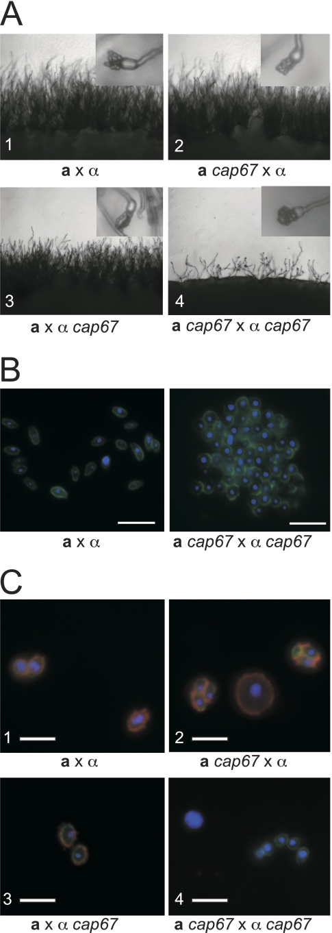
Capsule mutants have defects in sexual development and spore formation. (A) All panels show the periphery of a cross on V8 medium under a light microscope at low magnification (×200). Insets each show a single basidium under higher magnification (×400). (1) Wild-type a × α cross. (2 and 3) Wild-type a or α strain crossed with corresponding a cap67 or α cap67 strain. (4) a cap67 × α cap67 cross. The few basidia that were produced did not develop proper spore chains and yielded clumps of spores (inset in panel 4). Identical results were obtained for all additional capsule deletion strains tested (cap10Δ, cap60Δ, and cap64Δ strains). (B) Spores were stained with DAPI (blue) and DSL (green) and visualized by fluorescence micros- copy. (Left) Normal morphology of spores derived from a wild-type cross. (Right) Spores derived from a cap67 × cap67 cross forming an aggregated mass of spores. (C) Spores from wild-type and cap67 strains stained with DAPI to reveal nuclei (blue), with anti-GXM antibody (F12D2) to detect capsule (red), and with DSL to identify spores (green) were visualized by immunofluorescence microscopy. All three color channels were merged into a single image for each sample. (1) Spores derived from a wild-type a by α cross (JEC20 × JEC21). (2 and 3) Spores and yeast cells from crosses between wild-type and cap67 strains (CHY1377 × JEC21 and JEC20 × ATCC 52817, respectively). (4) Spores and yeast cells isolated from a cross between two cap67 strains (CHY1377 × ATCC 52817). Bars, 5 μm.

