FIG. 2.
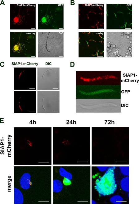
Expression and localization of PbSIAP-1/mCherry in sporozoites. Expression of the mCherry-tagged SIAP-1 (red) was analyzed by confocal fluorescence microscopy of SIAP-1/mCherry P. berghei parasites constitutively expressing GFP (green). (A) Midgut sporozoites. Bar, 20 μm. (B) Salivary gland-associated sporozoites. Bar, 10 μm. (C) Fluorescent imaging of a motile sporozoite. Sequential acquisition of the red fluorescence and differential interference contrast (DIC) confirms SIAP-1 accumulation mainly at the apical tip. Note that the distribution of SIAP-1/mCherry is not modified as the sporozoite glides. Bar, 5 μm. (D) Higher magnification of a fixed salivary gland-associated sporozoite. (E) Confocal microscopy analysis of HepG2 cell cultures 4 h, 24 h, and 72 h after infection with P. berghei parasites expressing GFP (green) and SIAP-1/mCherry (red). Nuclei were stained with Hoechst 33342 (blue). Bars, 10 μm.

