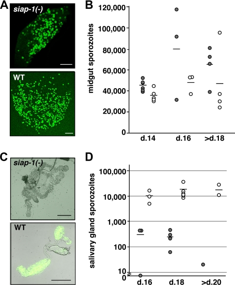
FIG. 4.
siap-1(−) sporozoites are impaired in egress from oocysts. (A) Representative immunofluorescence pictures of infected A. stephensi midguts at day 14 after infection. (B) Quantification of oocyst-associated sporozoites per infected mosquito for siap-1(−) (gray circles) and WT (white circles) parasites. The bars represent the mean values. (C) Representative merged bright field and immunofluorescence pictures of infected A. stephensi salivary glands at day 17 after infection. (D) Quantification of salivary gland-associated sporozoites per infected mosquito for siap-1(−) (gray circles) and WT (white circles) parasites. Note the logarithmic scale. Bars, 200 μm.

