FIG. 6.
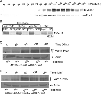
PAK function is required for Vac17-ProA degradation in late M phase. (A) VAC17-ProA PDS1-HA (YCH5162) cells were arrested in G1 with α-factor and released, samples were taken every 10 min, and Vac17-ProA and Pds1-HA were examined by Western blot analysis. (B) Vac17-ProA levels were assayed by Western blotting in cdc15-2 (YCH4862), dbf2-2 (YCH4869), and cdc14-1 (YCH4852) cells grown at 23°C or arrested at telophase in 37°C medium. (C and D) 4XGAL-CLA4t VAC17-ProA (YCH4894) and 4XGAL-CLA4t swe1 VAC17-ProA (YCH4893) cells were α-factor arrested and released, and samples were taken at the indicated times and subjected to Western blot analysis. Additionally, the percentage of telophase cells was determined by DAPI staining and the number of cells with mother and bud nuclei staining was scored.

