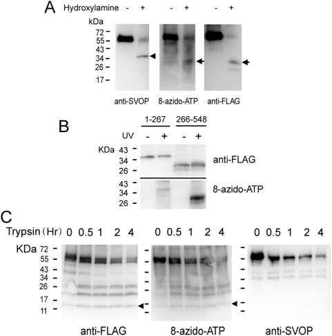
Figure 5. The nucleotide binding site in SVOP localizes to the C-terminal half of the protein.
A. 8-azido-ATP photoaffinity labeled SVOP-FLAG was cleaved by hydroxylamine and the samples were subjected to western blot analysis using anti-FLAG, anti-SVOP and ExtrAvidin-HRP. Arrowhead indicated an N-terminal fragment which is recognized by anti-SVOP antibody but not labeled by 8-azido-ATP. The Arrows indicate a C-terminal fragment which is labeled by 8-azido-ATP and anti-FLAG antibody. B. Only the C-terminal half of SVOP shows dominant nucleotide binding. N- and C-terminal halves of SVOP-FLAG were expressed and purified from HEK293 cells. Photoaffinity labeling was performed as described under Methods . C. Shown are the results of trypsin digestion of 8-azido-ATP labeled SVOP-FLAG. Labeled protein was digested at 37°C in the presence of trypsin. At the time periods indicated, an aliquot was withdrawn and subjected to analysis as described under Methods . The arrowheads indicate the smallest trypsinized 15 kDa fragment which is labeled by both 8-azido-ATP and anti-FLAG antibody. The proportion of total anti-FLAG and 8-azido-ATP labeling (undigested protein) was similar for the 15 KDa fragment (∼9% in both cases), consistent with it being labeled with the same efficiency as the full-length protein. Therefore the nucleotide-binding site is contained within this 15 KDa fragment. The image of 8-azido-ATP labeling blot was adjusted with contrast to show better results.

