
FIGURE 1.
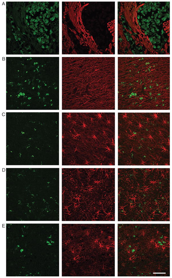
Microglial activation and reactive astrocytosis in various CNS disorders. Activated microglia (CD68, green) and reactive astrocytosis (GFAP, green) were assessed in various CNS diseases. Samples included the core of an acute cerebral infarct core (A), an active multiple sclerosis plaque (B), frontal cortex in a case of frontotemporal dementia (C), primary motor cortex in a case of amyotrophic lateral sclerosis (D), and frontal cortex in a case of Alzheimer disease (E). Individual (left and middle panels) and merged images (right panels) are shown. Scale bar = 50 μm.