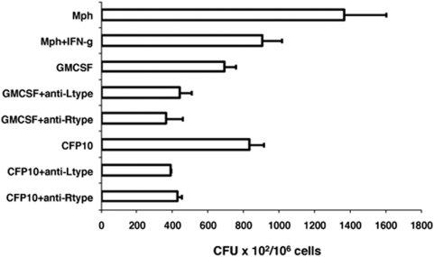
Figure 6. T cells activated by L-type and R-type VGCC-blocked-DCs kill M. tuberculosis inside macrophages.
L-type or R-type VGCC-blocked-BCG-infected GM-CSF-DCs (GMCSF) or CFP10-DCs (CFP10) were co-cultured with T cells from BCG immunized mice. From the co-culture, T cells were enriched and incubated with M. tuberculosis H37Rv infected macrophages. Cells were lysed and CFU was determined. As controls, infected macrophages without incubation with T cells and macrophages treated with 2 ng/ml IFN-γ prior to infection with M. tuberculosis in the absence of T cell addition were also included. Data are the mean of three independent experiments. Error bars represent mean±s.d. P<0.01 for Mph+IFN-g vs CFP10+anti-Ltype; P<0.01 for Mph+IFN-g vs CFP10+anti-Rype and P<0.006 for Mph vs GMCSF+anti-Ltype. Two-tailed Student's t-test was employed for P values.

