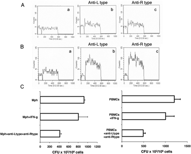
Figure 7. Blocking L-type and R-type VGCC in macrophages and PBMCs increases calcium influx and kills intracellular M. tuberculosis.
Real time increase in intracellular calcium over 5 min following stimulation of mouse macrophages (A) or human PBMCs (B) with 1 MOI BCG. Subpanel a- BCG stimulation, b & c anti-L-type and anti-R-type antibody blocking prior to BCG stimulation. C, M. tuberculosis H37Rv CFU in macrophages (left panel) or PBMCs (right panel) in the presence or absence of anti-L-type and anti-R-type antibody. IFN-g, cells incubated with 2 ng/ml IFN-γ prior to infection with M. tuberculosis H37Rv. Data are the mean of three independent experiments. Error bars represent mean±s.d. In C left panel P<0.09 for Mph+IFN-g vs Mph+anti-Ltype+anti-Rtype, P<0.005 for Mph vs Mph+anti-Ltype+anti-Rtype; right panel P<0.03 for PBMCs vs PBMCs+anti-Ltype+anti-Rtype, P<0.09 for PBMCs+IFN-g vs PBMCs+anti-Ltype+anti-Rtype. Two-tailed Student's t-test was employed for P values.

