
Fig. 2.
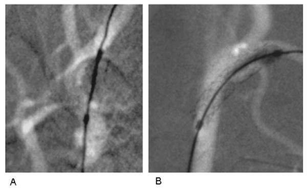
Fluoroscopic road-mapped frames done at 74 kVp and 50 mA of 3 mm diameter and 15 mm long coronary stent systems (Multi-link Vision, Guidant Corp, Santa Clara, CA). End markers indicate extent of expanded balloon on delivery catheter. Stents are deployed in carotid arteries of the same rabbit. (A) Using 2 ms pulsed XII imaging with temporal filtering (standard on commercial systems), stent deployed in right carotid. (B) Using 8 ms pulsed HSMAF imaging with no temporal filtering, stent deployed in left carotid showing improved visualization of details of stent struts and vessel edges.