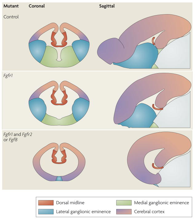
Figure 3. FGF signalling as an organizer for the telencephalon.
Schematics of coronal and sagittal sections in the telencephalon between embryonic day (E) 12.5 and E15.5 in various mutant mice. The colours represent areas that contain cells with different telencephalic identities. Progressively deleting more fibroblast growth factor receptor (FGFR) genes specifically in the anterior neural plate leads to diminished FGF signalling and gradually more severe truncations of telencephalic regions. Anterior ventral regions are lost first, followed by posterior dorsal regions34,38,46.

