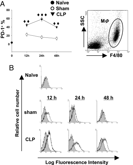
Fig. 2.
Expression of PD-1 in macrophages is augmented by sepsis. (A) The percentage of PD-1+ macrophage increased during sepsis. PD-1 expression on peritoneal macrophages (gated as F4/80+) was determined by flow cytometry (n = 5–12 for sham-treated mice, n = 7–17 for CLP-treated mice pooled from two to three experiments). ♦♦, P < 0.01, ♦♦♦, P < 0.001 by Mann-Whitney test, CLP-treated WT mice vs. sham-treated WT mice. (B) Representative histograms of PD-1 expression on peritoneal macrophages. Peritoneal macrophages were gated as F4/80+ peritoneal leukocytes (Upper). PD-1 expression (black lines) was overlayed on isotype control (gray filled; Lower).

