Fig. 1.
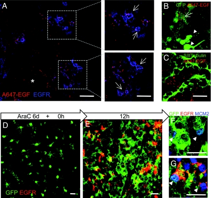
EGFR+ SVZ astrocytes are activated stem cells. (A–C) Validation of the use of EGF ligand to isolate EGFR-expressing SVZ cells. (A) In vivo binding of A647-EGF after intraventricular injection. Whole-mount preparation of the SVZ shows A647-EGF (red) bound to EGFR+ cells (blue). (Scale bar, 30 μm.) (Right) Higher-magnification view of boxes at Left. (Scale bar, 20 μm.) Note A647-EGF is also bound to noncellular extracellular matrix (asterisk). (B) Whole-mount preparation showing that A647-EGF binds to a subset of GFP dim cells (arrow) but not GFP bright cells (arrowhead) in GFAP::GFP mice. (C) Chains of neuroblasts (βIII tubulin, green) were not labeled by A647-EGF. (Scale bar for B, C, 20 μm.) (D–G) Analysis of SVZ astrocytes during regeneration at 0 (D) and 12 h (E) after AraC treatment with cytosine arabinofuranoside (AraC). (D) AraC treatment eliminates all GFP+EGFR+ cells, and only GFP+ (EGFR−) cells remain at 0 h. GFP+EGFR+ cells appear by 12 h (E). Both GFP+ (F) and GFP+EGFR+ (G) cells were labeled with anti-MCM2 (blue) at 12 h. GFP (green), EGFR (red). (Scale bars, 20 μm.)

