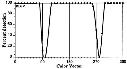
Figure 4.
Percentage of detection of 0.1° spots of different color vectors briefly presented in the central foveola. If S cones are missing within a small area of the central foveola, very small foveal stimuli of color vectors near 90° and 270° (where the L and M cone contrasts fall to zero but S cone contrast is maximal; see Fig. 1B) should be invisible, as they are for this observer.

