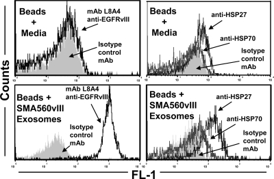
Figure 4.
Brain tumor exosome surfaces display EGFRvIII and heat shock proteins 27 and 70. Exosomes were bound to aldehyde-sulfate latex beads, were stained with fluorescently labeled antibodies to EGFRvIII (mAb L8A4; left panels) and to HSPs 27 and 70 (right panels), and were analyzed by flow cytometry. Staining of beads soaked in cell culture media as controls are shown in top panels. Isotype control staining profiles are shown as gray fill.

