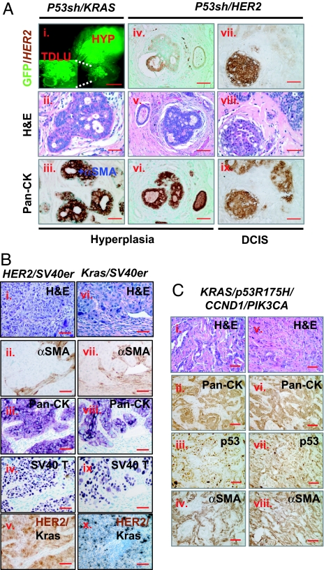
Fig. 1.
Human preneoplastic lesions and advanced breast cancers were generated in vivo from genetically engineered human breast tissue recombinants. (A) Premalignant lesions and CIS developed in vivo from human breast tissue recombinants overexpressing HER2 or KRAS with concomitant knocking down of p53. (Ai) GFP whole mount of reconstituted p53sh/KRAS/GFP human breast tissue reveals KRAS-lentivirus expression in both normal Terminal Ductal Lobular Unit (TDLU) structures (Inset) as well as in hyperplasic nodule (HYP). (Aii) H&E of the p53sh/KRAS/GFP hyperplastic outgrowth in Ai. (Aiii). IHC-stained serial section of Aii demonstrating the filling of the luminal space with epithelial cells. (Aiv–Aix) Histological analysis of p53sh/HER2 tissue recombinants revealed both hyperplastic (Aiv–Avi) and carcinoma in situ outgrowth (Avii–Aix) from transduced organoids (Aiv and Avii). (Scale bars: Ai, 0.5 mm; Aii–Aix, 50 μm.) (B) Poorly differentiated, invasive human carcinomas developed in vivo from HER2/SV40er and KRAS/SV40er human breast tissue recombinants. Histological analysis of HER2/SV40er (Bi–Bv) and KRAS/SV40er (Bvi–Bx) tumors. H&E-stained sections (Bi and Bvi) and IHC analysis on serial sections with αSMA (Bii and Bvii), pan-cytokeratin (Pan-CK; Biii and Bviii), SV40 LT (Biv and Bix), and HER2 (Bv) demonstrated that tumors were derived from epithelial cells that overexpressed the transduced oncogenes. RNA in situ analysis (Bx) confirmed the expression of KRAS in KRAS/SV40er tumors. (Scale bar: 100 μm.) (C) Invasive ductal adenocarcinoma developed in vivo from KRAS/p53R175H/CCND1/PIK3CA tissue recombinants. H&E-stained sections of genetically engineered HIM tumor (Ci and Cii) revealed that both samples were invasive ductal adenocarcinoma with prominent glandular architecture and low mitotic index. IHC-stained serial sections of HIM tumors with pan-cytokeratin (Pan-CK; Cii and Cvi), p53 (Ciii and Cvii), and αSMA (Civ and Cviii) revealed mutant p53-positive epithelial cancer cells surrounded by myofibroblastic stroma. (Scale bar: 50 μm.)

