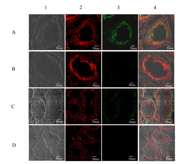
Figure 6.
Immunofluorescence staining of RNase9 in epididymis and testis. A, B: epididymis tissue; C, D: testis tissue; B, D: negative control. 1: phase contrast image; 2: nuclear counterstain (PI); 3: Green immunofluorescence staining of human RNase9 (FITC-labeled); 4: Merged image. Scale bar: 50 μm

