
Figure 4.
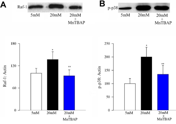
Effect of MnTBAP on glucose-induced activation of Raf-1 and phosphorylation of p38 MAP kinase. Activation of Raf-1 and phosphorylation of p38 MAP kinase were determined by Western blot using β-actin as a loading standard. Each sample was run in duplicate, and the experiment was repeated with three or more cell preparations. The histogram represents the density of Raf-1 (A), or p-p38 (B) band that has been adjusted to the density of β-actin band in the same lane. The ratio obtained from 5 mM glucose is considered as 100%. Asterisk (*) represent p<0.05 compared to untransfected cells in 5 mM glucose or MnSOD transfected cells incubated in 20 mM glucose, and double asterisk (**) marks p<0.05 compared to untransfected cells incubated in 20 mM glucose medium.