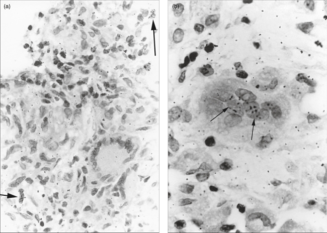
Fig. 3.

(a) Light microscopic appearance of a sarcoidosis lymph node section labelled with [3H]-thymidine. In the epithelioid cell granuloma two radio-labelled mononuclear cells (←) are seen, while a Langhans giant cell showed no radio-labelling [haematoxylin and eosin staining (H&E), ×400]. (b) In a granuloma section labelled with [3H]-uridine, a foreign body type giant cell is clearly visible in which some nuclei are radio-labelled (←) (H&E staining, ×1000).
