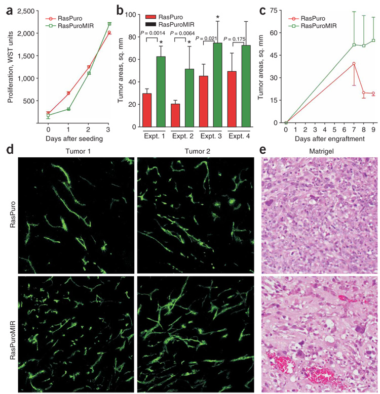
Figure 4. The effects of miR-17-92 upregulation in Ras-only cells on neoplastic growth.
(a) Cell accumulation assay performed on RasPuro and RasPuroMIR colonocytes. Numbers of viable cells were assessed using the WST reagent as in Figure 1a. (b) Average sizes of subcutaneous tumors formed by RasPuro and RasPuroMIR colonocytes in syngeneic mice. * indicates statistical significance (P < 0.05). P values were determined using unpaired Student’s t-test. (c) Kinetics of tumor formation by RasPuro and RasPuroMIR colonocytes from experiment 2 in b. Error bars in a–c represent s.d. (d) Blood perfusion of RasPuro and RasPuroMIR tumors. Green staining corresponds to FITC-conjugated lectin bound to vascular endothelial cells after intravenous injection. Two independent tumors were assayed. Representative 10× sections from each neoplasm are shown. (e) Matrigel neovascularization induced by the same cells. The richly perfused, large-caliber vascular channels in the lower image are typical of RasPuroMIR samples.

