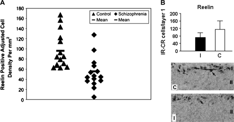
Fig. 3.
Reelin is Reduced in Hippocampus of Individuals With Schizophrenia and in Cerebral Cortex Following Prenatal Viral Infection. A. The values expressed on the y-axis are reelin-positive adjusted cell densities per square millimeter localized to hippocampal CA4 areas in control and schizophrenic subjects. The number of brains used is 15 (control) and 15 (schizophrenic). Each point is the mean for 2–4 sections analyzed per brain. A crossbar localized over each scatter plot represents mean Reelin-positive adjusted cell density value per group. Mean values for schizophrenic subjects are significantly reduced when compared with control values (analysis of variance, P < .05). B. The top panel shows a graph depicting the hemispheric Reelin-positive Cajal-Retzius (CR) cell counts in layer I of the cortex of prenatally infected (I) and sham-infected control (C) animals. The number of Reelin-positive CR cells was significantly reduced in infected brains compared with control brains (P < .0001). The lower panel shows light micrographs of layer I–II in coronal sections of prenatally infected and sham-infected cortex. Originally published in Fatemi et al.67,179

