
Figure 2.
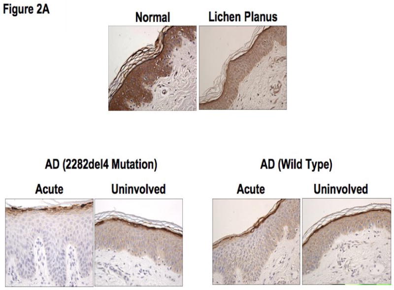
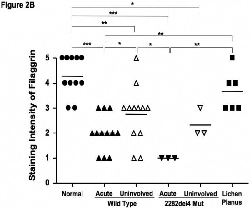
Decreased filaggrin staining in AD skin. A. Representative paraffin embedded skin biopsies from normal subjects (n=11), AD patients (n=12), AD patients with the 2282del4 mutation (n=3), and patients with lichen planus (n=6) stained for filaggrin are shown. Images were collected at 40× magnification and the scale bar represents 50 μm. B. The intensity of the staining was graded visually on a scale from 0 (no staining) to 5 (the most intense staining). *, **, and *** indicate significant differences of p<0.05, p<0.01, and p<0.001, respectively.