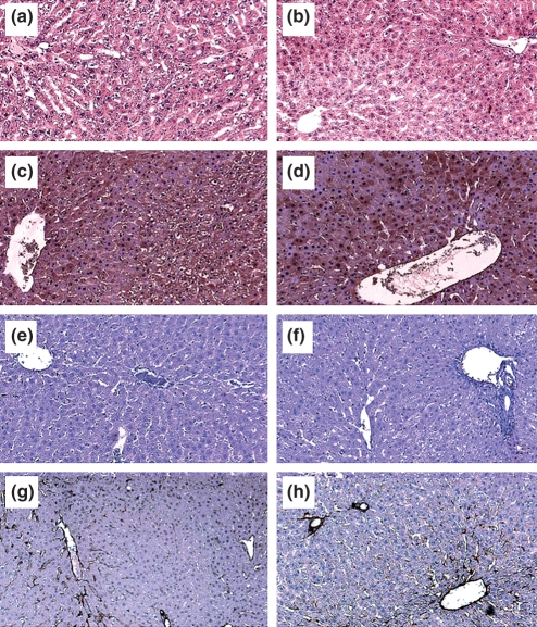
Figure 4.
Light microscopy and immunohistochemistry of livers from control and pyocyanin groups. Light microscopy of liver sections stained with eosin and haematoxylin staining. (a) Control liver, (b) pyocyanin-treated liver. Malondialdehyde immunohistochemistry [(c) control liver, (d) pyocyanin-treated liver], 3-nitrotyrosine immunohistochemistry [(e) control liver, (f) pyocyanin-treated liver] and caveolin-1 immunohistochemistry [(g) control liver, (h) pyocyanin-treated liver] revealed no changes across the hepatic lobule with pyocyanin treatment. Immunohistochemical stains appear brown, original magnification 100×.

