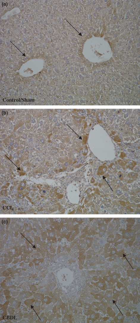
Figure 5.
VEGF immunohistochemistry (objective magnification 20×). (a) In control and sham-operated mice, VEGF was detected in the first layer of hepatocytes around the venules (black arrows). (b) After CCl4 and CBDL induction, the per centage hepatocytes that was positive increased with the highest concentration at 16 weeks for CCl4 administration (b) and at 3 weeks for CDBL induction (c).

