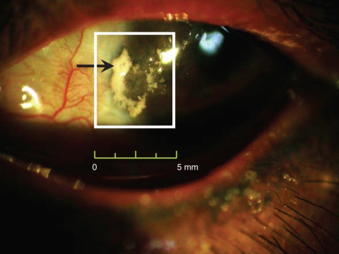
Figure 1.
A photograph of white calcium deposition in the sclera of patient with dystrophic calcification. The gross photography of this patient's left eye was taken by the digital camera equipped on the slit lamp microscope in the outpatient section of Ophthalmology Department. The size of calcified plaque was about 4.5 mm × 4.0 mm.

