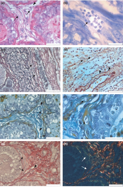
Figure 1.
Histopathological analysis of Swiss mice testis, 14 days after infection with Trypanosoma cruzi, CL strain. (a) Nests of T. cruzi amastigotes (arrows) are observed in the intertubular connective tissue near the blood–testis barrier. Dissociation of collagen fibres near seminiferous tubers are also seen (arrow head), (haematoxylin–eosin); (b) nests of T. cruzi are observed within myoid cells (arrow) (Lennert's Giemsa); (c) histochemical analysis for fibres with silver affinity puts in evidence an intimate relationship between type III collagen and parasite nests (arrow) (Gordon and Sweet's methods contrasted with eosin); (d) intertubular connective tissue in which are seen a T. cruzi nest (arrow) and non-modified elastic fibres (arrow head) (Weigert's resorcin–fucsin after oxone, oxidation counterstained with orange G); (e) intertubular connective tissue presenting collagen dissociation (arrow head) and parasitized myoid cell (arrow) (modified Shorr); (f) amastigote nest in a myoid cell cytoplasm (arrow) (modified Shorr); (g) intertubular connective tissue presenting parasite nest (arrow) and type I collagen dissociation (arrow head) (Picrosirius Red, non-polarized light), (h) intertubular connective tissue showing type III collagen supporting the parasite nest (green) (arrow), type I typical collagen (orange and red) and neoformed type I collagen (yellow) (Picrosirius Red, polarized light). All scale bars = 50 μm, except for B and F in which scale bars = 20 μm.

