
Figure 4.
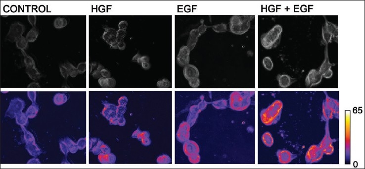
Increased membrane ruffling observed with HGF and EGF. H1993 were plated in glass bottom dishes coated with collagen (150,000 cells/plate). They were grown in serum-free medium containing 0.5% BSA for 24 h after which each plate was pretreated for 2 h with 100 ng/ml EGF, HGF or both. Cells were studied under TLVM for 4 h and photographs were taken at 1-min intervals. Cell movement with changing shape was analyzed using NIH image analysis. Images in the lowerpanel were created with false colors according to the movement and change in shape of cells. An increase in membrane ruffling was seen by an increase in red color, and a decrease is indicated by presence of blue color as observed in the bar on the side of Figure 4. Increased membrane ruffling was seen when cells were incubated with HGF or EGF and an additive effect was observed by a combination of both