
Figure 2.
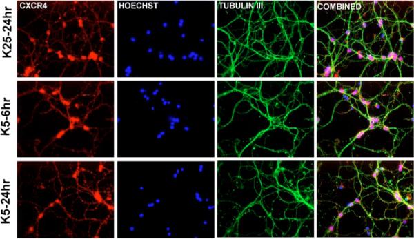
Expression of CXCR4 in cerebellar granule neurons cultured in high or low potassium. Control neurons (K25) and neurons maintained in K5 for 6 to 24 h were immunostained with a polyclonal antibody against CXCR4 (amino acids 176 to 293) and a monoclonal antibody for the specific neuronal marker μ-tubulin III (green). Nuclei were stained with Hoechst 33342 (blue).