
Fig. 5.
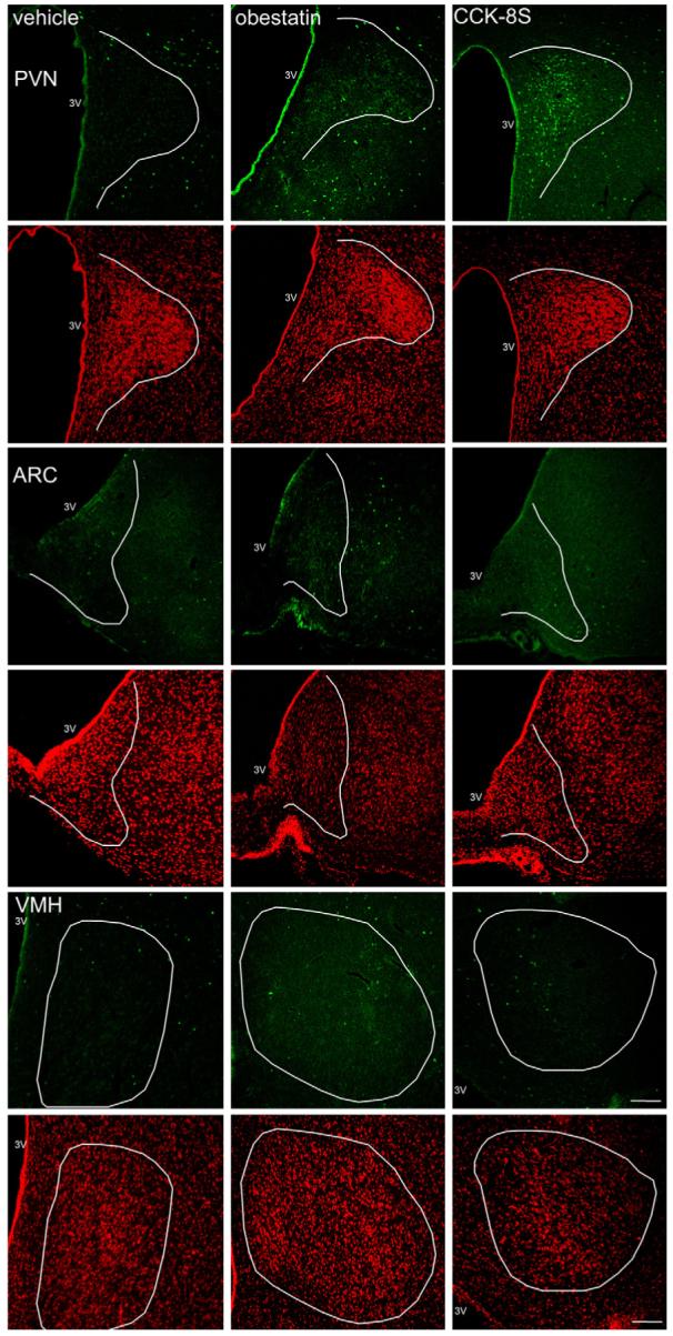
Representative images of the PVN, ARC and VMH after ip-injection of 0.15 M NaCl, 5 μmol/kg obestatin and 1.75 nmol/kg CCK-8S. Obestatin does not change neuronal activity in these hypothalamic nuclei while CCK-8S induces increased Fos expression (green staining) in the PVN. Cell nuclei are stained red as a result of the counterstaining with propidium iodide. The white outer line delineates the area of the PVN, ARC and VMH. The white scale bar represents 100 μm. 3V, third ventricle; PVN, paraventricular nucleus of the hypothalamus; ARC, arcuate nucleus of the hypothalamus; VMH, ventromedial nucleus of the hypothalamus. (For interpretation of the references to color in this figure legend, the reader is referred to the web version of the article.)