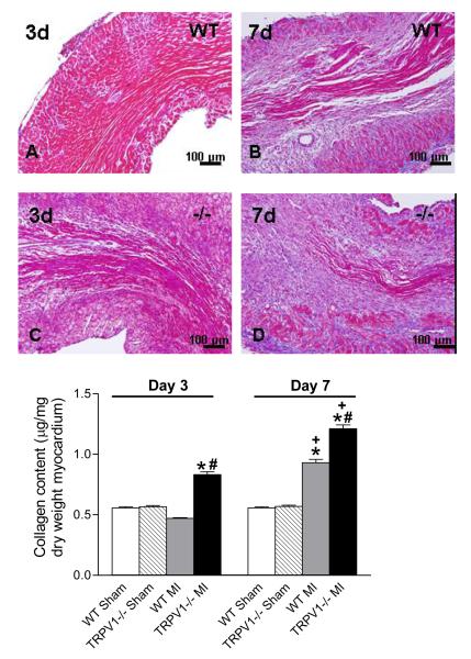
Figure 9.
Collagen deposition in the peri-infarct zone. Upper panel: Masson’s trichrome staining for collagen in the infarct region (X200, blue-stained). Lower panel: Quantitative collagen in various groups. N=8. *P<0.01 vs corresponding sham; †P<0.05 vs. WT at the same time point; ‡P<0.05 vs. corresponding day 3.

