
Fig. 3.
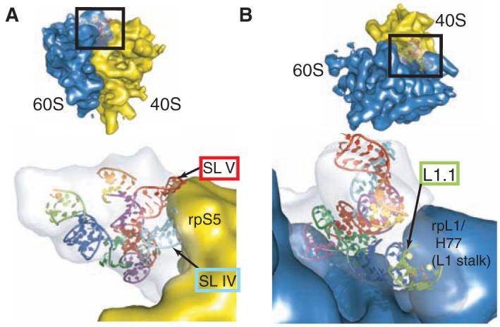
Interaction of the IGR IRES RNA with the ribosome. (A) The PSIV IGR IRES ribosome affinity domain structure docked into the cryo-EM representation of the IRES bound to the 80S ribosome, with the 60S ribosome density computationally removed. The 40S subunit is in yellow, the cryo-EM density of the IRES is in gray, and the IRES crystal structure is colored as in Fig. 1. The positions of rpS5 (40S subunit) and SL IV and SL V (IGR IRES) are shown. (B) Detailed view of the interaction of the IGR IRES to the 60S subunit within the 80S ribosome-IRES complex, with the 40S subunit density computationally removed. The L1 stalk of the 60S subunit contacts IRES loop L1.1 and perhaps P1.1. For both (A) and (B), the orientation is indicated in the insets.