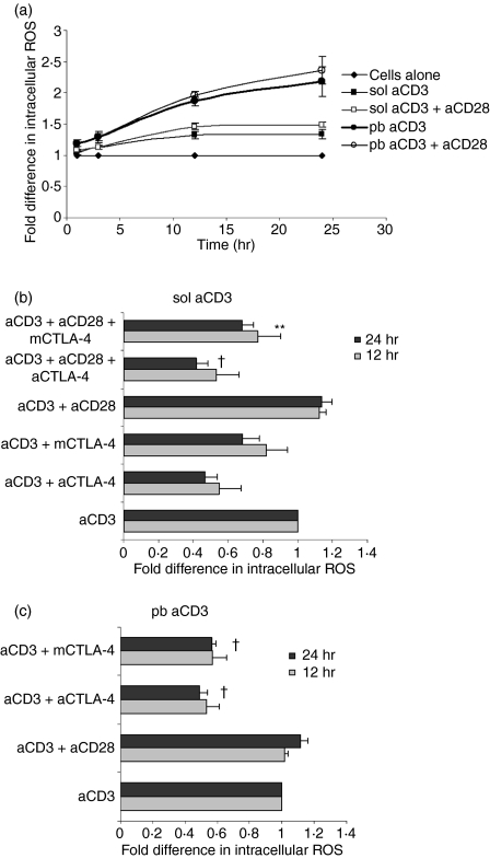
Figure 7.
Intracellular reactive oxygen species (ROS) increases with signal strength and cytotoxic T-lymphocyte antigen (CTLA)-4–CD80/CD86 interactions. Amounts of ROS were measured by 2′, 7′-dichlorofluorescein diacetate (DCFDA) fluorescence conversion at different times in cells activated with soluble anti-CD3 (sol aCD3) and plate-bound anti-CD3 (pb aCD3) ± anti-CD28. Mean fluorescence intensities (MFIs) under different conditions were normalized to the MFI of unactivated cells, which was taken as unity (a). A similar experiment was performed with blockade of CTLA-4–CD80/CD86 interactions and ROS was measured after 12 and 24 hr of activation. MFIs were normalized to that of cells activated with anti-CD3, which was taken as unity (b and c). Data shown are mean ± standard error for three independent experiments. †P< 0·0005; **P< 0·05.

