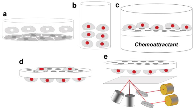
Figure 1.
Scheme outlining a novel method to quantify cell migration with near-infrared fluorescence. (a) Primary mouse microglial cells were harvested from mixed glial cultures by shaking. (b) Microglia were resuspended in serum-free MEM and stained with DRAQ5 (c) Stained cells were loaded in upper wells of a 96-well Boyden Chamber, with chemoattractant solutions in the lower wells. The chamber was then incubated for three hours, allowing cells to migrate toward the chemoattractant. (d) After incubation, filter was removed from the chamber and non-migrated cells on the top of the filter were wiped off. The filter was then fixed in methanol. (e) Fluorescence signal emitted by cells that did migrate was scanned using a LI-COR Odyssey scanner.

