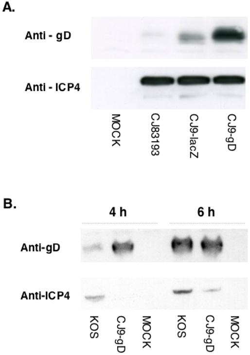
Figure 2. High-level expression of gD following CJ9-gD infection of Vero cells.

A.Vero cells were seeded at 1 × 10⁶ cells per 60-mm dish. At 23 h after seeding, cells in duplicate were either mock-infected or infected with CJ83193, CJ9-lacZ, or CJ9-gD at an MOI of 5 PFU/cell. Infected cell extracts were prepared at 24 h post-infection. B. Vero cells were infected with the wild-type HSV-1 strain KOS, or CJ9-gD at an MOI of 20 PFU/cell. Infected cell extracts were prepared at 4 h and 6 h post-infection. Proteins in infected cell extracts were resolved on SDS-PAGE, followed by immunoblotting with a monoclonal antibody specific for ICP4 or a polyclonal antibody specific for gD.
