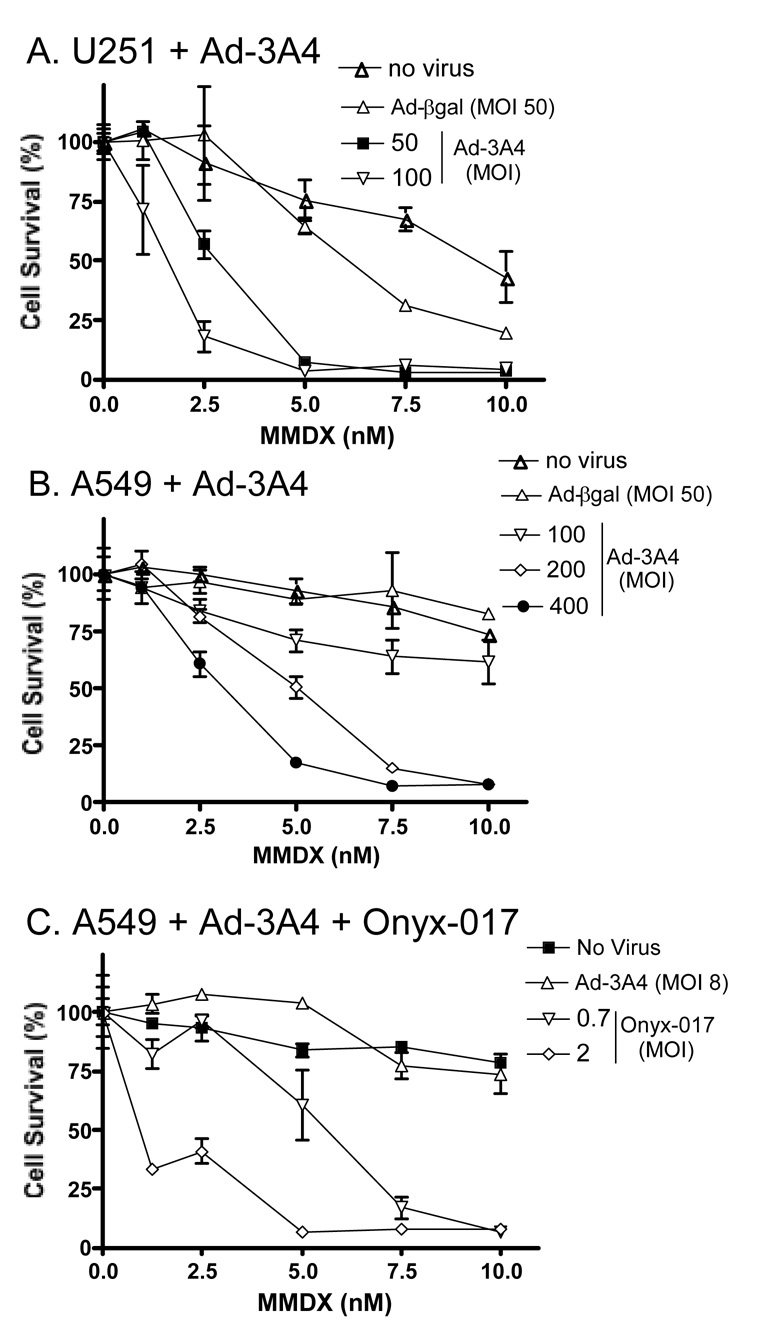
Fig. 3. Cytotoxicity of MMDX toward Adeno-3A4-infected U251 (A) and A549 cells (B, C), without (A, B) or with (C) Onyx-017 co-infection.
Cells seeded overnight in 24-well plates (14,000 cells/well) were infected with Adeno-3A4 or Adeno-βgal at the indicated MOI for 24 hr, either alone (A, B) or in combination with Onyx-017 (C; at MOIs 0.7 and 2). Cells were treated with MMDX in fresh culture medium at the indicated concentrations beginning 24 hr after infection. The culture medium was replaced with medium containing fresh MMDX 2 d later to minimize the intrinsic toxicity of the virus, and the incubation was continued for an additional 5 d (total of 7 d MMDX treatment). Data are expressed as percent cell survival compared to the corresponding drug-free controls, determined by crystal violet staining, mean ± SD (n=3). In the absence of MMDX, Adeno-3A4 was moderately toxic to U251 cells (≤30% cell killing at MOI 50 and 100) but not to A549 cells (<16% killing at MOI 200 and 400) (data not shown). The IC50 (MMDX) in A549 cells was 1 nM at 8 MOI Adeno-3A4 + 2 MOI Onyx-017 vs. no effect in A459 cells infected with 8 MOI Adeno-3A4 alone.

