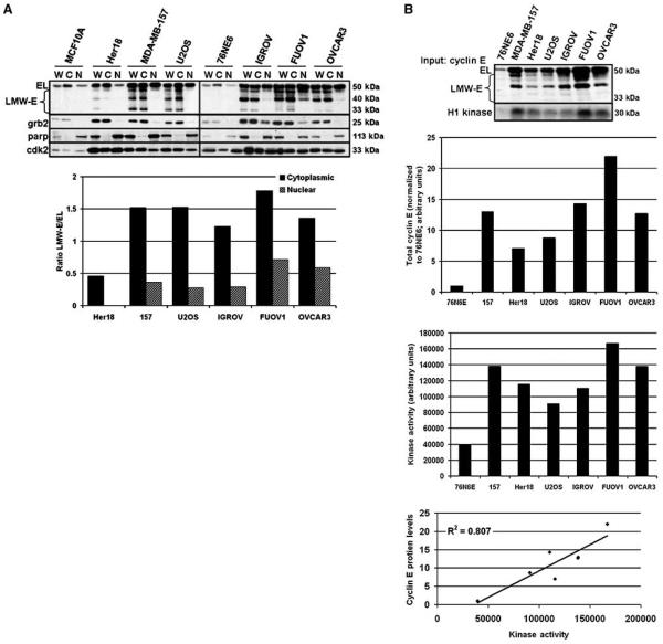
Figure 1. LMW-E isoforms are cytoplasmic.
A, normal immortalized mammary epithelial (MCF10A, 76NE6) cell lines and breast (Her18, MDA-MB-157), osteosarcoma (U2OS), and ovarian (IGROV, FUOV1, OVCAR3) cancer cell lines were separated into whole cell (W), cytoplasmic (C), and nuclear (N) fractions and analyzed for cyclin E and Cdk2 accumulation by Western blot. Grb2 and parp antibodies were used to indicate the cytoplasmic and nuclear fractions, respectively. Western blots were subject to densitometry and the ratio of total LMW-E to EL was graphed. B, the Western blot shows the relative levels of cytoplasmic cyclin E (Input: cyclin E) in each cell line that was assayed for cyclin E–associated kinase activity using histone H1 as a substrate (H1 kinase). Bar graphs, densitometric values for total cytoplasmic cyclin E (top bar graph; normalized to 76N6E) and H1 phosphorylation (bottom bar graph; raw values). Linear regression graph, correlation between cyclin E levels and cyclin E–associated kinase activity for each cell line; R2 = 0.807; P = 0.0060.

