
Figure 1.
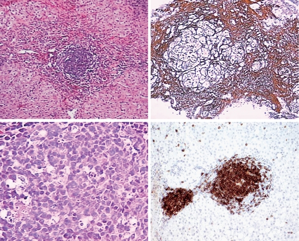
Histological appearances of a B-cell lymphoma, best classified as a marginal zone lymphoma involving the liver. A, B: Low power view of liver core biopsy shows chronic hepatitis and marked portal lymphoid infiltrates. C: High power view of liver core biopsy shows a monotonous portal lymphoid infiltrate composed of small lymphocytes with moderate amount of clear cytoplasm (so-called monocytoid appearance). Note that the infiltrate does not involve the biliary epithelium. D: CD20 immunostain shows that virtually all of the lymphoid cells are B-cells.