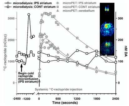
Fig. 3.
Dual-probe microdialysis measures of ECF DA with simultaneous microPET assay of 11C-raclopride time activity following a cold raclopride infusion into the ipsilateral striatum. Circles depict the time activity of 11C-raclopride (open) and extracellular DA (filled) in the striatum ipsilateral to an infusion of 45 nmol raclopride (corrected for probe permeability). Squares represent 11C-raclopride binding (open) and extracellular DA (filled) in the contralateral, control striatum. Triangles give the time activity of cerebellar 11C-raclopride. Time-activity data were fit to a tri-exponential model as described in the text. Inset: Top image captures 11C-raclopride microinfusion (∼50 μCi/probe) immediately following the study, the middle image depicts the distribution of 11C-raclopride at the level of the striatum. The two images are co-registered with PMOD software on the bottom. Right is right in the image.

