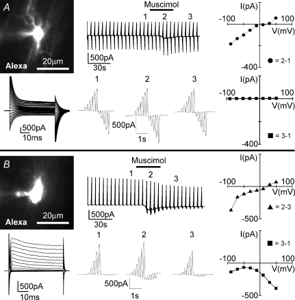
Figure 6. Muscimol-induced current response from an oligodendrocyte and a glial progenitor cell in the corpus callosum.
An oligodendrocyte with characteristic tail current (A) and a glial progenitor cell with voltage-gated membrane current (B) in the neighbourhood of amoeboid microglia in the postnatal corpus callosum were voltage-clamped. Images of cells filled with Alexa Fluor 594 (10 μg ml−1) via patch pipette are shown (top left). Membrane currents (top middle and bottom left) were recorded as described in Fig. 1 in response to muscimol (100 μm; 30 s). Voltage steps before (1), during (2) and after (3) muscimol-induced response are shown at a higher time resolution (bottom middle). Current–voltage plots were obtained by subtracting current responses at various time points as indicated (right).

